Comprehensive Library Of Resveratrol News

Subscribe to our newsletter to receive email notifications when new articles are posted.


  • A Tale Of Two Sirtuins

    June 30, 2011: by Bill Sardi

    Has The Holy Grail Of Anti-Aging Been (Re-)Discovered?

    KEY POINTS

    • Thepromise of the Sirtuin1 gene as anti-aging target has been disappointing.
    • Long-living humans found to have active Sirtuin3 gene.
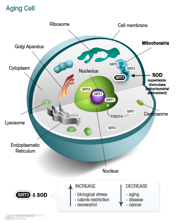
    • Sirtuin1 gene located in cell cytoplasm whereas Sirtuin3 controls antioxidant protection (SOD*) inside mitochondria where cell energy and 90% destructive oxygen free radicals are produced.
    • Feverishly-paced research is underway to confirm Sirtuin3 as true anti-aging pill.
    • Resveratrol molecule stimulates Sirtuin3 gene; Longevinex® resveratrol matrix activates Sirtuin3 2.95-fold greater than plain resveratrol.

    * SOD = superoxide dismutase

    Genes they be — a Sirtuin family of seven “silent information regulators” that are guardians of the cell. Sirtuins have been linked with prolonged lifespan in various life forms, starting with yeast cells, fruit flies and roundworms.1

    Sirtuin1 (also referred to as SIRT1) is a survival gene that is switched on when a living organism is deprived of food. Since the lifespan of calorie-restricted laboratory animals nearly doubles, researchers thought, for sure, they had struck biological pay dirt. Researchers thought any molecule that could turn on Sirtuin1 (trigger it to make proteins) could be an anti-aging molecule for humans. In fact, one Harvard genetics professor claimed Sirtuin1 was the “holy grail” of anti-aging2 and demonstrated in the laboratory that it could be activated by a red wine molecule called resveratrol.

    All this was first announced in Nature magazine in late 20033, was extolled on the front pages of The New York Times4 and The Wall Street Journal5, and followed by a laboratory experiment which showed resveratrol prolonged the life of fat-fed mice6, and by 2010 a biotech company sold their experimental resveratrol-based drug to a major pharmaceutical company for $720 million.7

    From promise to disappointment

    But that wasn’t quite how it all ended. A later study showed mega-dose resveratrol slightly shortened the lifespan of laboratory rats fed a standard-calorie diet, though it did dramatically improve other measurable health parameters largely by eradicating fatty liver.8

    Then an MIT professor chimed in and said the pursuit of longevity via Sirtuin1 was a bit more complicated than first thought and that a calorie restricted diet didn’t uniformly up-regulate the Sirtuin1 gene in all organs and tissues.9 Dratted science never seems to cooperate just when humanity needed such a pill.

    Then more confusion — the initial experiment which showed resveratrol activated Sirtuin1 was flawed. Turns out a fluorescent dye used in gene analysis foiled the experiment as it was found to be the agent that switched on Sirtuin1, not resveratrol.10 That didn’t negate the incomparable health benefits attributed to resveratrol, but it did negate Sirtuin1 as its primary gene target. Sounds like the pharmaceutical company ought to be asking for its money back. The major pharmaceutical company did announce it was not continuing further research and development of the SRT501 drug. Did the drug company remove SRT501 from its R&D pipeline to make sure it doesn’t compete with today’s disappointing prescription drugs.

    Two genes away: Sirtuin3

    But a month before the 2003 Nature magazine paper was published about Sirtuin1, researchers in Italy published an overlooked report concerning genetic data from a pool of humans over 100 years of age. These researchers found a relationship exists between longevity and genes located near the 11p15.5 chromosome. Five genes potentially involved in longevity were located and scrutinized. In this chromosomal region lies Sirtuin3, a gene that resides inside the energy power plants inside cells called mitochondria, whereas Sirtuin1 resides in the watery cytoplasm inside living cells.

    A significant variation in survivorship was found among those individuals who exhibited a form of Sirtuin3. To date, among all seven Sirtuin genes, it is the only one genetically linked to lifespan in humans.11 But at the time, this discovery was overshadowed by the excitement for Sirtuin1.

    Fast forward in time to 2011 – the science pointing to Sirtuin3 as a true anti-aging gene is mounting rapidly. As one research paper described it: “Sirtuin3 has recently stepped out of the shadow of Sirtuin1… mimicking calorie restriction.” Sirtuin3 was found to increase cellular respiration by 80% compared to 30% for Sirtuin1.12 Sirtuin1 gene’s own pied piper, Harvard researcher David Sinclair, filed for a patent in 2008 claiming activation of Sirtuin3 mimics the metabolic effect of endurance exercise training.13 No one noticed.

    The whole tenor of anti-aging researchers has changed from the confusion and disappointment experienced with Sirtuin1. Usually reserved scientists are now saying things like “Sirtuin3 may be a central mechanism of aging retardation in mammals,14 “Sirtuin3 may have profound implications for aging,15 and that “a barrage of recent studies show that Sirtuin3 wards off the vicissitudes of aging.16

    To further corroborate the link between premature aging and lack of Sirtuin3 proteins, researchers have found that Sirtuin3 activity declines with advancing age.17

    Surprisingly, if the Sirtuin3 gene is removed from laboratory mice, no overt signs of distress are observed. But if these same Sirtuin3-absent mice are subjected to biological or mental stress (food deprivation, excessive radiation, etc.), these stressed mice produce heightened levels of free radicals and decreased levels of cellular energy (ATP – adeno-triphosphate).18 Sirtuin3 is the antidote to harmful species of oxygen molecules known as free radicals, which are missing a key electron.

    Why is Sirtuin3 superior to Sirtuin1?

    Why does Sirtuin3 appear to work against aging in a superior manner to Sirtuin1? The answer to this question may lie in the cellular compartment where it is found – the mitochondria. Sirtuin 3, 4 and 5 are found within the hundreds of mitochondrial compartments within living cells, which is where cellular respiration (conversion of oxygen to energy) takes place and where 90% of potentially destructive oxygen free radicals in the body are produced.

    Sirtuin3 controls the internal (endogenous) antioxidant response to free radicals in the most critical of places – the mitochondria. It increases the activity of a natural endogenous antioxidant enzyme called superoxide dismutase (SOD) in the mitochondria.

    The mitochondria are where 85-95% of oxygen used by cells is consumed to produce cellular energy and where over 90% of the destructive oxygen free radicals (oxidizing agents) are generated. By age 80 only about 4% of mitochondria are actually functioning at full capacity.19 This fact alone should indicate why Sirtuin3 may have such a profound and promising role as a single gene out of a library of about 25,000 genes. While according to one recent study human aging involves at least 295 genes20, Sirtuin3 appears to single-handedly counter the biological chaos that occurs within the mitochondria.

    Think of a living cell with an outer fatty membrane holding in the watery cytoplasm. Within that cell is a nucleus that houses the genetic material – DNA. (See diagram below.) In the watery cytoplasm are hundreds of atomic power plants (mitochondria) that generate cell energy via respiration of oxygen. About 5% of that oxygen turns into potentially harmful oxygen free radicals, what is called oxidation or rusting. It’s like fires are burning in the hundreds of mitochondria in the cell with advancing age which must be extinguished by the manganese form of SOD via control by the Sirtuin3 gene. Put those mitochondrial fires out and cells in the brain, heart, liver, live longer and better withstand biological stress.

    These facts do not negate the fact that the proper form of SOD (manganese-SOD) is also activated by resveratrol via the Sirtuin1 gene pathway.21 However, Sirtuin1 does not reside in the critical location that Sirtuin3 does.

    Here are other known facts about this strategically placed gene:

    • Sirtuin3 proteins are found in a broad range of tissues and are consistently increased with biological stress, unlike Sirtuin1.
    • Sirtuin3 has been shown to increase cellular energy (ATP).22
    • Sirtuin3 is the master gene that controls the production of SOD in the mitochondria. A deficiency of SOD is associated with many diseases, particularly diseases of aging.23
    • Sirtuin3 is considered a tumor suppressor by virtue of its ability to activate SOD within the mitochondria.22
    • Sirtuin3 proteins are also increased by calorie restriction.24
    • An age-related decline in Sirtuin3 has now been associated with heart failure.25
    • Sirtuin3 also activates catalase, another endogenous antioxidant enzyme.26
    • A large percentage of human tumors show inactivation of Sirtuin3.27
    • The precursor for SIRT3-generated SOD is the metallic mineral manganese. With the absence or shortage of manganese, iron may take its place.28
    • Unbound iron that can generate destructive free radicals can enter the mitochondria.29
    • In mice lacking the Sirtuin3 gene, they rapidly develop a form of breast cancer that has been linked with a low SOD activity levels.30
    • Resveratrol, a red wine molecule, has been shown to increase manganese-SOD activity.31 Longevinex® is the first branded resveratrol dietary supplement demonstrated to activate Sirtuin3.32 Comparatively, Longevinex® appears to activate Sirtuin3 nearly three-fold greater than plain resveratrol (1.18 to 0.4 arbitrary units).
    Comparative Activation Of Sirtuin3 Gene
    Regulator of Mitochondrial SOD Antioxidant Activity
    Arbitrary Units Fold Increase
    Resveratrol 0.40
    Longevinex® resveratrol-based matrix 1.18 +2.95 (295%)
    • Quercetin33, ferulic acid34 and IP635 (derived from rice bran), provided in the Longevinex® formula, are also known to be protective antioxidants in the mitochondria.
    • Sirtuin3 is known to reverse the age-related conversion in the generation of cellular energy from oxygen burning during youth to sugar burning in the latter years of life. Sirtuin3 also helps to maintain normal levels of oxygen in the mitochondria necessary to inhibit activation of the hypoxia inducing factor gene (HIF1). Tumor cells grow in a hypoxic (oxygen-starved) environment. The activation of Sirtuin3 reverses the Warburg effect in breast cancer cells growing in lab dishes. Two-time Nobel Prize winner Otto Warburg was the first to describe the phenomenon in the 1930s, where tumor cells convert from using oxygen to utilizing sugar to survive.36

    Sirtuin3 and hearing loss

    An intriguing animal study was conducted among mice that have been bred for hearing loss. If these animals are given a calorie restricted diet, despite their genetic predisposition to develop hearing loss, this sparse calorie diet completely prevented this occurrence. However, if the Sirtuin3 gene is removed from these animals and then they are given a limited calorie diet, the protection against hearing loss is completely negated.37

    Corroboration of the beneficial effects of Longevinex® in humans have been received by researchers who have documented that older males experience hearing improvement as measured by audiological examination.38

    What now?

    Researchers are working feverishly on Sirtuin3 gene research. Will unfolding research result in a re-run of what occurred in 2003 with all the public fascination spawned by widespread news media coverage that surrounded resveratrol as a Sirtuin1 gene activator? Will the news media continue to ignore the stellar performance of the Longevinex® nutriceutical, which has many times now outperformed the $720 million SRT501 resveratrol drug?39,40,41,42 Will the National Institute on Aging launch a study to confirm whether resveratrol or a resveratrol-based matrix actually prolongs the life of laboratory animals? Stay tuned.
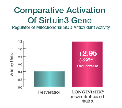
    Comparison: Serituin 3 activation


    Aging Cell


    More than just SOD

    While manganese-SOD may be a primary protector of the mitochondria, it only converts troublesome destructive oxygen molecules called free radicals into hydrogen peroxide (H2O2), which itself is potentially destructive. Another internally-produced antioxidant enzyme, catalase, must convert hydrogen peroxide (H2O2) into harmless water (H2O). The process is O2 (reactive oxygen) -> H2O2 (hydrogen peroxide) + catalase -> = H2O (water).


    Now if you read up on catalase, you would think it was the key antioxidant that produces longevity. In fact, in 2005 Science magazine published a report entitled “The Anti-Aging Sweepstakes: Catalase Runs for the ROSes” (ROS = reactive oxygen species).43

    And there is strikingly convincing evidence for catalase as a key anti-aging antioxidant. For example, among mice bred to over-produce catalase, median lifespan was maximally increased by 5.0 to 5.5 months (these lab animals only live 2.0-2.5 years, so this is a 16-24% jump that in human terms would be equivalent to another 15-20 years of healthy living).44 Understand, that is five times greater predicted life extension than what can be achieved by elimination of cancer and heart disease in these animals. In mice bred to over-produce catalase, all of the age-related changes in heart function were significantly reduced.45

    Catalase probably deserves as much or more attention if for nothing else than one molecule of catalase can break 40 million molecules of hydrogen peroxide every second, turning it to harmless water.
    Also consider that while other antioxidants play a role in the mitochondria, they are often not able to deal with the large amount of oxidation (reactive oxygen species) and the mitochondria often lack sufficient catalase activity.46 With -38% chemically-induced reduction of catalase, normal cells display accelerated aging. The mitochondria become dysfunctional.47

    When the human catalase gene is inserted in laboratory mice, they live longer, which is even more of an enticement to jump to the conclusion that catalase is THE longevity molecule.48

    The unheralded endogenous antioxidants

    While consumers hear of the many protective properties of dietary and supplemental antioxidants such as vitamin C, vitamin E, coenzyme Q10 and others, three primary enzymatic antioxidants (catalase, superoxide dismutase and glutathione peroxidase) produced in living cells are overlooked.

    Indeed, both selenium and vitamin E, the precursors for glutathione peroxidase, are required to protect mitochondria from damage by hydrogen peroxide.49

    When researchers conducted a study to measure free radical production in the brains of different vertebrate animals, they found that SOD, catalase, glutathione peroxidase, the three major internally-produced antioxidants, along with vitamin C, correlated with maximum longevity. Low levels of free radical production and low antioxidant concentrations are a central finding in long-lived animals.50

    Contrary data

    However, there is contrary data that confounds a clear oxidant-antioxidant explanation of aging. High levels of oxidative damage in mitochondrial DNA do not decrease longevity in mice.51 Contrary to other experiments, when researchers intentionally induced overproduction of SOD, catalase, or combinations of the two, it didn’t result in extended lifespan of mice.52 In another mouse study, life-long reduction of MnSOD activity led to increased levels of oxidative damage to DNA and increased cancer incidence but did not appear to affect aging.53

    Yet with all the praise for catalase, some researchers say the majority of studies using antioxidant strategies “do not support a clear role for mitochondrial oxidative stress or a vicious cycle of oxidative damage in the determination of lifespan in mice and furthermore do not support the free radical theory of aging.”54

    In mice, the provision of increasing amounts of supplemental coenzyme Q10, a widely promoted antioxidant supplement, did not raise SOD, catalase or glutathione peroxidase antioxidant levels in the mitochondria and had no effect upon mortality.55

    Of course, there is nothing better than human studies to test the antioxidant theory of aging. So recently researchers at the US Dept of Agriculture Human Nutrition Research Center on Aging at Tufts University enrolled 46 moderately overweight volunteers, age 20-42 years, and were given diets of differing reduced calorie load. Only glutathione peroxidase levels increased over a 6-month period of caloric restriction. No other antioxidants such as SOD or catalase were altered in blood plasma.56

    Still, while scientific controversy reigns, researchers at the Division of Geriatrics, University of California at Los Angeles believe we are on the cusp of a breakthrough in the pursuit of human longevity and state that “the advancement of mitochondrially-targeted small-molecule antioxidants suggests the prospect of swift translation to human use.”57

    King resveratrol

    Certainly, resveratrol rises to the top of a list of candidates as anti-aging molecules, with good reason. Examine these studies:In a lab dish, at very low concentration, resveratrol increases activity of the three major antioxidant enzymes, SOD, catalase and glutathione peroxidase, in cells obtained from the human retina.58 This occurs without provision of the nutrients (copper, zinc, manganese, vitamin E, selenium) necessary to produce these antioxidants.

    It has been clearly demonstrated that resveratrol activates all three key antioxidant enzymes (glutathione, SOD and catalase) in arteries.59

    Does resveratrol always shine in the laboratory? Well, in one experiment, at very low concentration, resveratrol had no effect upon key antioxidant enzymes such as catalase, glutathione peroxidase or copper-zinc variety of SOD in the lab dish. But, as researchers described it in their own words: “resveratrol dramatically and progressively induced mitochondrial manganese-SOD activity.” Two weeks into their experiment resveratrol increased manganese-SOD protein levels by 6-fold and Mn-SOD activity levels by 14-fold!60

    Test tube studies however are not real live environments. So researchers put resveratrol to the test in mice, in what are called in vivo experiments. The lab mice were fed a standard calorie diet, a high-fat diet and were also fed via an osmotic pump to ensure the feeding was the same between animals. Unsurprisingly, resveratrol increased manganese-SOD protein-making by 140% and its activity by 75%.61

    Is aging too large for biology to get its hands around it?

    When one considers all of the factors that come into play in aging it would appear to be a daunting challenge to develop an anti-aging substance that would address all of the genes and biological processes involved, ranging from inflammation and oxidation to DNA breaks and enzyme control. But then again, there is resveratrol. Just examine how researchers explain the breadth of resveratrol’s biological influence:

    Extensive research within the last decade has revealed that most chronic illnesses such as cancer, cardiovascular and pulmonary diseases, neurological diseases, diabetes, and autoimmune diseases exhibit dysregulation of multiple cell signaling pathways that have been linked to inflammation. Thus mono-targeted therapies developed for the last two decades for these diseases have proven to be unsafe, ineffective and expensive. Resveratrol, a polyphenol, has been shown to mediate its effects through modulation of many different pathways. This molecule has been shown to bind to numerous cell-signaling molecules such as multi drug resistance protein, topoisomerase II, aromatase, DNA polymerase, estrogen receptors, tubulin and F1-ATPase. Resveratrol has also been shown to activate various transcription factor (e.g; NFkappaB, STAT3, HIF-1alpha, beta-catenin and PPAR-gamma), suppress the expression of antiapoptotic gene products (e.g; Bcl-2, Bcl-X(L), XIAP and survivin), inhibit protein kinases (e.g; src, PI3K, JNK, and AKT), induce antioxidant enzymes (e,g; catalase, superoxide dismutase and hemoxygenase-1), suppress the expression of inflammatory biomarkers (e.g., TNF, COX-2, iNOS, and CRP), inhibit the expression of angiogenic and metastatic gene products (e.g., MMPs, VEGF, cathepsin D, and ICAM-1), and modulate cell cycle regulatory genes (e.g., p53, Rb, PTEN, cyclins and CDKs). Numerous animal studies have demonstrated that this polyphenol holds promise against numerous age-associated diseases including cancer, diabetes, Alzheimer, cardiovascular and pulmonary diseases. In view of these studies, resveratrol’s prospects for use in the clinics are rapidly accelerating. Efforts are also underway to improve its activity in vivo through structural modification and reformulation. Our review describes various targets of resveratrol and their therapeutic potential.”62

    What causes age-related increase in mitochondrial oxidation?

    A question arises. If oxidation within the mitochondria increases with advancing age, and the mitochondria cease to function optimally in advanced age, what causes the progressive deterioration within the mitochondria? Genes do not run the show, they only respond to environmental cues within living cells.

    This author maintains the accumulation of minerals, namely copper, iron and calcium, is what controls the rate of aging. Indeed, a recent report clearly describes how potentially destructive metals like copper, iron, even chromium and cobalt, produce metal-induced free radicals such as superoxide and the dreaded hydroxyl radical which then overwhelm antioxidant systems and damage DNA.63

    Copper is the more powerful oxidant and it is of interest to note that while all metallic metals can induce potentially destructive free radicals, copper has been demonstrated to “overwhelm antioxidant defenses” (catalase, glutathione, SOD) more so than iron and other metallic minerals.64 As copper levels rise in the blood plasma, the risk for heart disease rises also.65 That cannot be said for iron. And resveratrol is “by far, the most potent chelator of copper.”66

    It is interesting to note that females live longer than males in many species, including humans. And the mitochondria from females produce significantly less hydrogen peroxide than those from males and have higher levels of mitochondrial reduced glutathione, manganese superoxide dismutase, and glutathione peroxidase than males. Oxidative damage to mitochondrial DNA is also fourfold higher in males than in females. Researchers indicate “these differences may be explained by estrogens. Surgical removal of estrogen-producing ovaries abolishes the gender differences between males and females and estrogen replacement rescues the ovariectomy effect. The challenge for the future is to find molecules that have the beneficial effects of estradiol, but without its feminizing effects. Phytoestrogens or phytoestrogen-related molecules may be good candidates to meet this challenge.”67Resveratrol is a safe phytoestrogen.

    Sorting it all out

    What are we learning here? The messages to take home are large and many for longevinarians.

    1. Sirtuin3 is probably primary among the family of seven Sirtuin genes, and may be a sole genetic marker for longevity, but it doesn’t work alone and obviously works within networks of survival/longevity genes and overlapping antioxidant systems.
    2. Antioxidant enzymes, that is, those internally (endogenously)-produced antioxidants may take precedence over externally provided antioxidants as provided in the diet (examples: vitamin C, vitamin E), but do require nutrient precursors, such as selenium + vitamin E = glutathione peroxidase; manganese-SOD = manganese; catalase = zinc, copper, manganese.
    3. Not only is Sirtuin3 involved, but it works in tandem with Sirtuin4 and Sirtuin1 and both influence another gene, FOXO1, to “translocate” from the watery cytoplasm to the nucleus of the cell. What signal triggers this is yet unknown. Additionally, Sirtuin6 is newly recognized as a required gene in double-strand DNA repair.68
    4. Then again, with all that has been learned up to this point, even low-grade superoxide radical challenge induces an adaptive state and triggers defenses in the body, which is beneficial (so-called hormetic action where a low-grade toxin is protective and high-grade toxin produces cell and possibly total organism death).
    5. There is even another unheralded antioxidant enzyme, peroxiredoxin (Prx) that detoxifies hydrogen peroxide in the mitochondria and is linked to longevity. So we can’t pin all of our hopes on one gene or a single antioxidant.
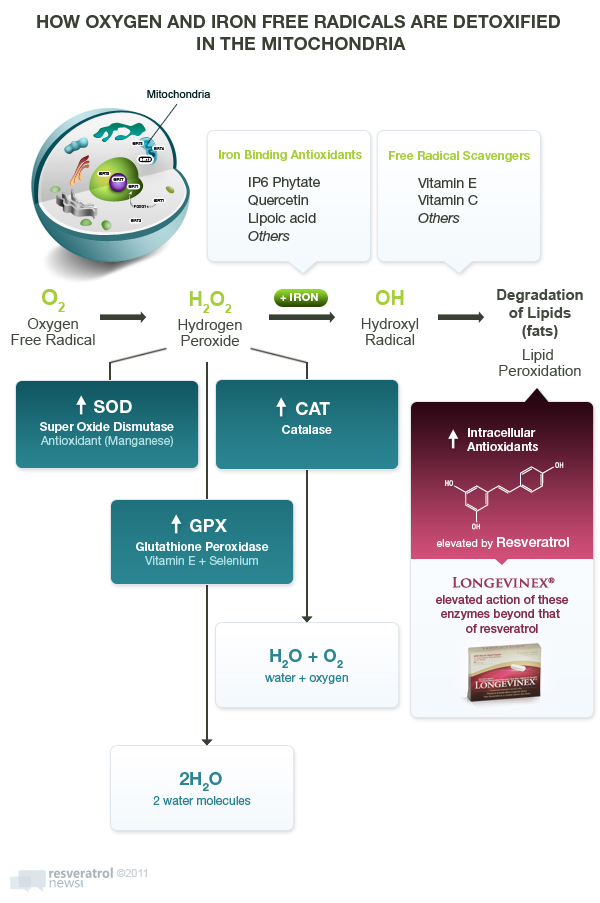
    6. Resveratrol, and more so Longevinex®, activates intracellular antioxidants within the mitochondria, without provision of their nutrient precursors (copper, manganese, zinc, selenium, vitamin E).


    free radicals detox in mitochondria


    Follow-up questions and answers:

    Question: the form of SOD (superoxide dismutase) antioxidant within the mitochondria is produced from manganese. Is adding manganese to the diet an anti-aging strategy?

    Reply: Here is what the manganese-SOD molecule looks like:
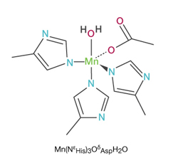
    manganese


    SOD is formed from three precursors – copper, zinc and manganese. Manganese-SOD constitutes approximately 10-15% of the total SOD activity in most tissues.

    Manganese SOD appears to be a more powerful antioxidant than the copper or zinc-SODs under biologically stressed conditions.69

    SOD activity is increased in response to biological stress. A frank deficiency of manganese and reduced manganese-SOD activity has been demonstrated to be deleterious to the heart. Provision of supplemental manganese has been shown to be therapeutic.70 However, the heart-protective properties of manganese were only demonstrated in manganese-deficient animals compared to manganese-sufficient animals.71

    Higher intake of manganese from the diet, but not necessarily supplements, is associated with lower risk for brain disease.72 Inorganically-bound manganese in drinking water is potentially more troublesome than bound forms of manganese in the diet.73 In animals, chronic exposure to manganese and other heavy metals in drinking water was counterproductive and actually decreased SOD activity over the long term.74

    Supplemental manganese has drawn cause for concern in multivitamins.75 The estimated safe daily intake for manganese is 2-5 mg per day, however dosage range for side effects begins at just 4.2 mg/day for a 70-kilogram/160-lb individual. So the potentially problematic dose falls within the so-called safe range.76

    A high intake of manganese when accompanied with iron is also of concern in relation to age-related brain disease.77 With advancing age, metallic metals accumulate in living tissues. The accumulation of manganese may be deleterious.78

    The Western diet appears to generally provide sufficient amounts of manganese.79

    Provision of oral SOD itself without biological stress may sometimes be problematic. Manganese is a heavy metal that is only needed in minute amounts to produce SOD. Excessive activation of manganese-SOD, while anticipated to be increased under biological stress, may not be beneficial when constantly increased when biological stress is low or absent.80

    Question: what about taking oral SOD itself?

    Reply: SOD is a antioxidant enzyme that is rapidly degraded by gastric acids when ingested orally. Even enteric coated tablets do not adequately protect SOD and SOD supplements are said to produce no pharmacologic activity when taken orally. Oral SOD was found to ineffectively raise SOD levels in laboratory mice.81 However, this is a dated study and only used low (less than 1mg) doses.

    Oral SOD may be of some value when administered directly into tissues. The provision of oral SOD directly into the gut reduced oxidative stress in animals with colitis. A 910-mg equivalent human dose (13 mg/kilogram for a 70 kilogram/160-lb human) was employed.82

    Various brands of oral SOD are sold in retail shops. The bioavailability of oral SOD is not equivalent to what is employed in the research laboratory where injectable SOD is often used. Oral SOD is poorly bioavailable unless special encapsulation methods are employed.83 Manganese-SOD has a high molecular weight of around 81,000 Daltons and would be expected to be poorly absorbed.84 For comparison, the molecular weight of readily absorbed vitamin C is 176 Daltons. Low molecular weight SOD has been successfully employed in animal studies, but was administered intravenously.85 The trick is to activate SOD in the mitochondria, not to assume that oral SOD makes its way to remote tissues beyond the digestive tract.

    References

    To access abstract, click on PubMed (National Library of Medicine) master identification number (PMID) at end of citation.

    1. Milne JC, Denu JM, The Sirtuin family: therapeutic targets to treat diseases of aging. Curr Opin Chem Biol. 2008 Feb;12(1):11-7. PMID:18282481
    2. The Associated Press, Big doses of red wine could promote long life, 11/1/2006
      http://www.msnbc.msn.com/id/15511128/ns/health-health_care/t/big-doses-red-wine-could-promote-long-life/
    3. Howitz KT, Bitterman KJ, Cohen HY, Lamming DW, Lavu S, Wood JG, Zipkin RE, Chung P, Kisielewski A, Zhang LL, Scherer B, Sinclair DA. Small molecule activators of sirtuins extend Saccharomyces cerevisiae lifespan. Nature. 2003 Sep 11; 425(6954):191-6. PMID: 12939617
    4. Nicholas Wade, New Hints Seen That Red Wine May Slow Aging. The New York Times, June 4, 2008 http://www.nytimes.com/2008/06/04/health/research/04aging.html
    5. David Stipp, Researchers seek key to anti-aging in calorie cutback. The Wall Street Journal Tuesday, October 31, 2006 http://www.post-gazette.com/pg/06304/734200-114.stm
    6. Baur JA, Pearson KJ, Price NL, Jamieson HA, Lerin C, Kalra A, Prabhu VV, Allard JS, Lopez-Lluch G, Lewis K, Pistell PJ, Poosala S, Becker KG, Boss O, Gwinn D, Wang M, Ramaswamy S, Fishbein KW, Spencer RG, Lakatta EG, Le Couteur D, Shaw RJ, Navas P, Puigserver P, Ingram DK, de Cabo R, Sinclair DA. Resveratrol improves health and survival of mice on a high-calorie diet. Nature. 2006 Nov 16;444(7117):337-42. PMID: 17086191
    7. GlaxoSmithKline to acquire Sirtris Pharmaceuticals, a world leader in ‘Sirtuin’ research and development. Tuesday 22 April 2008
      http://www.gsk.com/media/pressreleases/2008/2008_us_pressrelease_10038.htm
    8. Pearson KJ, Baur JA, Lewis KN, Peshkin L, Price NL, Labinskyy N, Swindell WR, Kamara D, Minor RK, Perez E, Jamieson HA, Zhang Y, Dunn SR, Sharma K, Pleshko N, Woollett LA, Csiszar A, Ikeno Y, Le Couteur D, Elliott PJ, Becker KG, Navas P, Ingram DK, Wolf NS, Ungvari Z, Sinclair DA, de Cabo R. Resveratrol delays age-related deterioration and mimics transcriptional aspects of dietary restriction without extending life span. Cell Metab. 2008 Aug;8(2):157-68. PMID: 18599363
    9. Chen D, Bruno J, Easlon E, Lin SJ, Cheng HL, Alt FW, Guarente L. Tissue-specific regulation of SIRT1 by calorie restriction. Genes Dev. 2008 Jul 1;22(13):1753-7. PMID: 18550784
    10. Kaeberlein M, McDonagh T, Heltweg B, Hixon J, Westman EA, Caldwell SD, Napper A, Curtis R, DiStefano PS, Fields S, Bedalov A, Kennedy BK. Substrate-specific activation of sirtuins by resveratrol. J Biol Chem. 2005 Apr 29;280(17):17038-45. PMID: 15684413
    11. Rose G, Dato S, Altomare K, Bellizzi D, Garasto S, Greco V, Passarino G, Feraco E, Mari V, Barbi C, BonaFe M, Franceschi C, Tan Q, Boiko S, Yashin AI, De Benedictis G. Variability of the SIRT3 gene, human silent information regulator Sir2 homologue, and survivorship in the elderly. Exp Gerontol. 2003 Oct;38(10):1065-70. PMID: 14580859
    12. Dransfeld CL, Alborzinia H, Wölfl S, Mahlknecht U. SIRT3 SNPs validation in 640 individuals, functional analyses and new insights into SIRT3 stability. Int J Oncol. 2010 Apr;36(4):955-60. PMID: 20198340
    13. USPTO: Patent application title: USE OF COMPOUNDS ACTIVATING SIRT-3 FOR MIMICKING EXERCISE Inventors: David A. Sinclair (Chestnut Hill, MA, US) Juan Carmona (Belton, TX, US) Assignees: President and Fellows of Harvard College
      IPC8 Class: AA61K317088FI USPC Class: 514 44 R
      Publication date: 04/07/2011 Patent application number: 20110082189 http://www.faqs.org/patents/app/20110082189
    14. Someya S, Yu W, Hallows WC, Xu J, Vann JM, Leeuwenburgh C, Tanokura M, Denu JM, Prolla TA. Sirt3 mediates reduction of oxidative damage and prevention of age-related hearing loss under caloric restriction. Cell. 2010 Nov 24;143(5):802-12. PMID: 21094524
    15. Bell EL, Guarente L, The SirT3 Divining Rod Points to Oxidative Stress. Mol Cell. 2011 Jun 10;42(5):561-8. PMID: 21658599
    16. Liu Y, Zhang D, Chen D, SIRT3: Striking at the heart of aging. Aging (Albany NY). 2011 Jan;3(1):1-2. PMID: 21094524
    17. Lanza IR, Short DK, Short KR, Raghavakaimal S, Basu R. Joyner MJ, McConnell JP, Nair KS, Endurance exercise as a countermeasure for aging. Diabetes.2008 Nov;57(11):2933-42. PMID: 21248372
    18. Schumacker PT. A tumor suppressor SIRTainty. Cancer Cell. 2010 Jan 19;17(1):5-6. Review. PMID: 20129243
    19. Balaban RS, Nemoto S, Finkel T. Mitochondria, oxidants, and aging. Cell. 2005 Feb 25;120(4):483-95. PMID: 15734681
    20. Harries LW, Human aging is characterized by focused changes in gene expression and deregulation of alternative splicing. Aging Cell June 13, 2011 PMID: 21668623
    21. Tanno M, Kuno A, Yano T, Miura T, Hisahara S, Ishikawa S, Shimamoto K, Horio Y, Induction of manganese superoxide dismutase by nuclear translocation and activation of SIRT1 promotes cell survival in chronic heart failure. J Biol Chem 285 (11): 8375-82, 2010. PMID: 20089851
    22. Ahn BH, Kim HS, Song S, Lee IH, Liu J, Vassilopoulos A, Deng CX, Finkel T. A role for the mitochondrial deacetylase Sirt3 in regulating energy homeostasis. Proc Natl Acad Sci U S A. 2008 Sep 23;105(38):14447-52. PMID: 18794531
    23. Chen Y, Zhang J, Lin Y, Lei Q, Guan KL, Zhao S, Xiong Y, Tumor suppressor SIRT3 deacetylase and activates manganese superoxide dismutase to scavenge ROS. EMBO Reports 12(6): 534-41, 2011. PMID: 21566644
    24. Hallows WC, Yu W, Smith BC, Devries MK, Ellinger JJ, Someya S, Shortreed MR, Prolla T, Markley JL, Smith LM, Zhao S, Guan KL, Denu JM. Sirt3 promotes the urea cycle and fatty acid oxidation during dietary restriction. Mol Cell. 2011 Jan 21;41(2):139-49. PMID: 21255725
    25. Sadoshima J Sirt3 targets mPTP and prevents aging in the heart. Aging (Albany NY). 2011 Jan;3(1):12-3. PMID: 21248376
    26. Schumacker PT, A tumor suppressor SIRTainty. Cancer Cell. 2010 Jan 19;17(1):5-6. PMID: 20129243
    27. Finley LW, Carracedo A. Lee J, et al, SIRT3 opposes reprogramming of cancer cell metabolism through HIF1a destabilization. Cancer Cell 19: 416-28, 2011. PMID: 21397863
    28. Naranuntarat A, Jensen LT, Pazicni S, Penner-Hahn JE, Culotta VC. The interaction of mitochondrial iron with manganese superoxide dismutase. J Biol Chem. 2009 Aug 21;284(34):22633-40. PMID: 19561359
    29. Breuer W, Shvartsman M, Cabantchik ZI, Intracellular labile iron. Int J Biochem Cell Biol. 2008;40(3):350-4. PMID: 17451993
    30. Hursting SD, Lavigne JA, Berrigan D, Perkins SN , Barrett JC. Calorie restriction, aging, and cancer prevention: mechanisms of action and applicability to humans. Annual Review of Medicine 54: 131-52, 2003. PMID: 12525670
    31. Valdecantos MP, Pérez-Matute P, Quintero P, Martínez JA, Vitamin C, resveratrol and lipoic acid actions on isolated rat liver mitochondria: all antioxidants but different. Redox Rep. 2010;15(5):207-16. PMID: 21062536
    32. Mukherjee S, Ray D, Lekli I, Bak I, Tosaki A, Das DK. Effects of Longevinex (modified resveratrol) on cardioprotection and its mechanisms of action. Can J Physiol Pharmacol. 2010 Nov;88(11):1017-25. PMID: 21076489
    33. Potenza L, Calcabrini C, De Bellis R, Mancini U, Cucchiarini L, Dachà M. Effect of quercetin on oxidative nuclear and mitochondrial DNA damage. Biofactors. 2008;33(1):33-48. PMID: 19276535
    34. Lin YH, Zhang JJ, Chen WW. Protective effect of sodium ferulate on damage of the rat liver mitochondria induced by oxygen free radicals. Yao Xue Xue Bao. 1994;29(3):171-5. PMID: 8079647
    35. Panduri V, Weitzman SA, Chandel NS, Kamp DW, Mitochondrial-derived free radicals mediate asbestos-induced alveolar epithelial cell apoptosis. Am J Physiol Lung Cell Mol Physiol. 2004 Jun;286(6):L1220-7. PMID: 14766669
    36. Bell EL, Guarente L, The SirT3 divining rod points to oxidative stress. Molecular Cell 42: 561-68, 2011. PMID: 21658599
    37. Lombard DB, Alt FW, Cheng HL, Bunkenborg J, Streeper RS, Mostoslavsky R, Kim J, Yancopoulos G, Valenzuela D, Murphy A, et al, Mallalian Sir2 homolog regulates global mitochondrial lysine acetylation. Mol Cell Biol 27, 8807-8814, 2007. PMID: 17923681
    38. Data received from Stuart Richer OD, PhD, Director, Ocular Preventive Medicine- Eye Clinic, James A Lovell Federal Health Care Center, North Chicago, Illinois.
    39. Juhasz B, Mukherjee S, Das DK, Hormetic response of resveratrol against cardioprotection. Exp Clin Cardiol. 2010 Winter;15(4):e134-8. PMID: 21264071
    40. Juhasz B, Das DK, Kertesz A, Juhasz A, Gesztelyi R, Varga B, Reduction of blood cholesterol and ischemic injury in the hypercholesteromic rabbits with modified resveratrol, longevinex. Mol Cell Biochem. 2011 Feb;348(1-2):199-203. PMID: 21052791
    41. Mukherjee S, Ray D, Lekli I, Bak I, Tosaki A, Das DK, Effects of Longevinex (modified resveratrol) on cardioprotection and its mechanisms of action. Can J Physiol Pharmacol. 2010 Nov;88(11):1017-25. PMID: 21076489
    42. Mukhopadhyay P, Mukherjee S, Ahsan K, Bagchi A, Pacher P, Das DK. Restoration of altered microRNA expression in the ischemic heart with resveratrol. PLoS One. 2010 Dec 23;5(12):e15705. PMID: 21203465
    43. Miller RA, Biomedicine. The anti-aging sweepstakes: catalase runs for the ROSes. Science. 2005 Jun 24;308(5730):1875-6. PMID: 15976292
    44. Schriner SE, Linford NJ, Martin GM, Treuting P, Ogburn CE, Emond M, Coskun PE, Ladiges W, Wolf N, Van Remmen H, Wallace DC, Rabinovitch PS.Extension of murine life span by overexpression of catalase targeted to mitochondria.Science. 2005 Jun 24;308(5730):1909-11. PMID: 15879174
    45. Dai DF, Santana LF, Vermulst M, Tomazela DM, Emond MJ, MacCoss MJ, Gollahon K, Martin GM, Loeb LA, Ladiges WC, Rabinovitch PS.Overexpression of catalase targeted to mitochondria attenuates murine cardiac aging. Circulation. 2009 Jun 2;119(21):2789-97. PMID: 19451351
    46. Bai J, Cederbaum AI. Mitochondrial catalase and oxidative injury. Biol Signals Recept. 2001 May-Aug;10(3-4):189-99.PMID: 11351128
    47. Koepke JI, Wood CS, Terlecky LJ, Walton PA, Terlecky SR. Progeric effects of catalase inactivation in human cells. Toxicol Appl Pharmacol. 2008 Oct 1;232(1):99-108. PMID: 18634817
    48. Enns LC, Wiley JC, Ladiges WC. Clinical relevance of transgenic mouse models for aging research. Crit Rev Eukaryot Gene Expr. 2008;18(1):81-91. PMID: 18197787
    49. Combs GF Jr, Noguchi T, Scott ML. Mechanisms of action of selenium and vitamin E in protection of biological membranes. Fed Proc. 1975 Oct;34(11):2090-5.
      PMID: 1100438
    50. Barja G, Cadenas S, Rojas C, López-Torres M, Pérez-Campo R. A decrease of free radical production near critical targets as a cause of maximum longevity in animals. Comp Biochem Physiol Biochem Mol Biol. 1994 Aug;108(4):501-12. PMID: 7953069
    51. Sanz A, Stefanatos RK. The mitochondrial free radical theory of aging: a critical view. Curr Aging Sci. 2008 Mar;1(1):10-21. PMID: 20021368
    52. Pérez VI, Van Remmen H, Bokov A, Epstein CJ, Vijg J, Richardson A. The overexpression of major antioxidant enzymes does not extend the lifespan of mice. Aging Cell.2009 Feb;8(1):73-5. PMID: 19077044
    53. Van Remmen H, Ikeno Y, Hamilton M, Pahlavani M, Wolf N, Thorpe SR, Alderson NL, Baynes JW, Epstein CJ, Huang TT, Nelson J, Strong R, Richardson A. Life-long reduction in MnSOD activity results in increased DNA damage and higher incidence of cancer but does not accelerate aging. Physiol Genomics. 2003 Dec 16;16(1):29-37.
      PMID: 14679299
    54. Jang YC, Remmen VH. The mitochondrial theory of aging: insight from transgenic and knockout mouse models. Exp Gerontol. 2009 Apr;44(4):256-60. PMID: 19171187
    55. Sohal RS, Kamzalov S, Sumien N, Ferguson M, Rebrin I, Heinrich KR, Forster MJ.
      Effect of coenzyme Q10 intake on endogenous coenzyme Q content, mitochondrial electron transport chain, antioxidative defenses, and life span of mice. Free Radic Biol Med. 2006 Feb 1;40(3):480-7. PMID: 16443163
    56. Meydani M, Das S, Band M, Epstein S, Roberts S. The Effect of Caloric Restriction and Glycemic Load on Measures of Oxidative Stress and Antioxidants in Humans: Results from the CALERIE Trial of Human Caloric Restriction. J Nutr Health Aging. 2011;15(6):456-60. PMID: 21623467
    57. Wanagat J, Dai DF, Rabinovitch P.Mitochondrial oxidative stress and mammalian healthspan. Mech Ageing Dev. 2010 Jul-Aug;131(7-8):527-35. PMID: 20566356
    58. Pintea A, Rugină D, Pop R, Bunea A, Socaciu C, Diehl HA. Antioxidant Effect of Trans-Resveratrol in Cultured Human Retinal Pigment Epithelial Cells. J Ocul Pharmacol Ther. 2011 Jun 11. PMID: 21663493
    59. Li H, Förstermann U. Resveratrol: a multifunctional compound improving endothelial function. Editorial to: “Resveratrol supplementation gender independently improves endothelial reactivity and suppresses superoxide production in healthy rats” by S. Soylemez et al. Cardiovasc Drugs Ther. 2009 Dec;23(6):425-9. PMID: 19937102
    60. Robb EL, Page MM, Wiens BE, Stuart JA. Molecular mechanisms of oxidative stress resistance induced by resveratrol: Specific and progressive induction of MnSOD. Biochem Biophys Res Commun. 2008 Mar 7;367(2):406-12. PMID: 18167310
    61. Robb EL, Winkelmolen L, Visanji N, Brotchie J, Stuart JA. Dietary resveratrol administration increases MnSOD expression and activity in mouse brain. Biochem Biophys Res Commun. 2008 Jul 18;372(1):254-9. PMID: 18486604
    62. Harikumar KB, Aggarwal BB. Resveratrol: a multitargeted agent for age-associated chronic diseases. Cell Cycle. 2008 Apr 15;7(8):1020-35. PMID: 18414053
    63. Jomova K, Valko M. Advances in metal-induced oxidative stress and human disease. Toxicology. 2011 May 10;283(2-3):65-87. PMID: 21414382
    64. Fahmy B, Cormier SA. Copper oxide nanoparticles induce oxidative stress and cytotoxicity in airway epithelial cells. Toxicol In Vitro. 2009 Oct;23(7):1365-71.
      PMID: 19699289
    65. Ford ES. Serum copper concentration and coronary heart disease among US adults. Am J Epidemiol. 2000 Jun 15;151(12):1182-8. PMID: 10905530
    66. Belguendouz L, Fremont L, Linard A. Resveratrol inhibits metal ion-dependent and independent peroxidation of porcine low-density lipoproteins. Biochem Pharmacol. 1997 May 9;53(9):1347-55. PMID: 9214696
    67. Viña J, Sastre J, Pallardó F, Borrás C. Mitochondrial theory of aging: importance to explain why females live longer than males. Antioxid Redox Signal. 2003 Oct;5(5):549-56. PMID: 14580309
    68. Mao Z, Hine C, Tian X, Van Meter M, Au M, Vaidya A, Seluanov A, Gorbunova V.SIRT6 promotes DNA repair under stress by activating PARP1. Science. 2011 Jun 17;332(6036):1443-6. PMID: 21680843
    69. Tsunoda R, Okumura K, Ishizaka H, Matsunaga T, Tabuchi T, Tayama S, Yasue H, Enhancement of myocardial reactive hyperemia with manganese-superoxide dismutase: role of endothelium-derived nitric oxide. Cardiovasc Res. 1996 Apr;31(4):537-45. PMID: 8689645
    70. Malecki EA, Greger JL, Manganese protects against heart mitochondrial lipid peroxidation in rats fed high levels of polyunsaturated fatty acids. J Nutr. 1996 Jan;126(1):27-33. PMID: 8558311
    71. Pucheu S, Coudray C, Tresallet N, Favier A, de Leiris J, Effect of dietary antioxidant trace element supply on cardiac tolerance to ischemia-reperfusion in the rat. J Mol Cell Cardiol. 1995 Oct;27(10):2303-14. PMID: 8576945
    72. Miyake Y, Tanaka K, Fukushima W, Sasaki S, Kiyohara C, Tsuboi Y, Yamada T, Oeda T, Miki , Kawamura N, Sakae N, Fukuyama H, Hirota Y, Nagai M; Fukuoka Kinki Parkinson’s Disease Study Group. Dietary intake of metals and risk of Parkinson’s disease: A case-control study in Japan.J Neurol Sci. 2011 Jul 15;306(1-2):98-102. PMID: 21497832
    73. Bouchard MF, Sauvé S, Barbeau B, Legrand M, Brodeur MÈ, Bouffard T, Limoges E, Bellinger DC, Mergler D, Intellectual impairment in school-age children exposed to manganese from drinking water. Environ Health Perspect. 2011 Jan;119(1):138-43. PMID: 20855239
    74. Jadhav SH, Sarkar SN, Aggarwal M, Tripathi HC, Induction of oxidative stress in erythrocytes of male rats subchronically exposed to a mixture of eight metals found as groundwater contaminants in different parts of India. Arch Environ Contam Toxicol. 2007 Jan;52(1):145-51. PMID: 17031751
    75. Catherine A Mulholland and Diane J Benford, What is known about the safety of multivitamin-multimineral supplements for the generally healthy population? Theoretical basis for harm.Am J Clin Nut, Vol. 85, No. 1, 318S-322S, January 2007 PMID: 17209218
    76. Greger JL, Dietary standards for manganese: overlap between nutritional and toxicological studies. J Nutr. 1998 Feb;128(2 Suppl):368S-371S. PMID: 9478027
    77. Powers KM, Smith-Weller T, Franklin GM, Longstreth WT Jr, Swanson PD, Checkoway H, Parkinson’s disease risks associated with dietary iron, manganese, and other nutrient intakes. Neurology. 2003 Jun 10;60(11):1761-6. PMID: 12796527
    78. The Manganese Health Research Program, Brief Background on the Health Effects of Manganese, http://www.manganese-health.org/about_us/healtheffects
    79. Greger JL, Nutrition versus toxicology of manganese in humans: evaluation of potential biomarkers. Neurotoxicology. 1999 Apr-Jun;20(2-3):205-12. PMID: 10385884
    80. Bödör C, Matolcsy A, Bernáth M, Elevated expression of Cu, Zn-SOD and Mn-SOD mRNA in inflamed dental pulp tissue. Int Endod J. 2007 Feb;40(2):128-32. PMID: 17229118
    81. Zidenberg-Cherr S, et al. Dietary superoxide dismutase does not affect tissue levels. Am J Clin Nutrit 1983;37:5. PMID: 6849282
    82. Seguí J, Gironella M, Sans M, Granell S, Gil F, Gimeno M, Coronel P, Piqué JM, Panés J. Superoxide dismutase ameliorates TNBS-induced colitis by reducing oxidative stress, adhesion molecule expression, and leukocyte recruitment into the inflamed intestine. J Leukoc Biol. 2004 Sep;76(3):537-44 PMID: 15197232
    83. Regnault C, Soursac M, Roch-Arveiller M, Postaire E, Hazebroucq G, Pharmacokinetics of superoxide dismutase in rats after oral administration. Biopharm Drug Dispos. 1996 Mar;17(2):165-74. PMID: 8907723
    84. Sugiura M, Adachi T, Inoue H, Ito Y, Hirano K, Purification and properties of two superoxide dismutases from human placenta. J Pharmacobiodyn. 1981 Apr;4(4):235-44.
      PMID: 7264869
    85. Lawrence de Garavilla, Timothy Chermak, Heather L. Valentine, and
      Robert C. Hanson, Novel Low-Molecular-Weight Superoxide Dismutase Mimic
      Deferoxamine-Manganese Improves Survival Following Hemorrhagic and
      Endotoxic Shock. Drug Development Research 25: 139-148 (1992)
      http://onlinelibrary.wiley.com/doi/10.1002/ddr.430250206/abstract

    Copyright 2011- Bill Sardi

Leave a Reply


In order to submit your comment please complete the calculation below:

Time limit is exhausted. Please reload CAPTCHA.